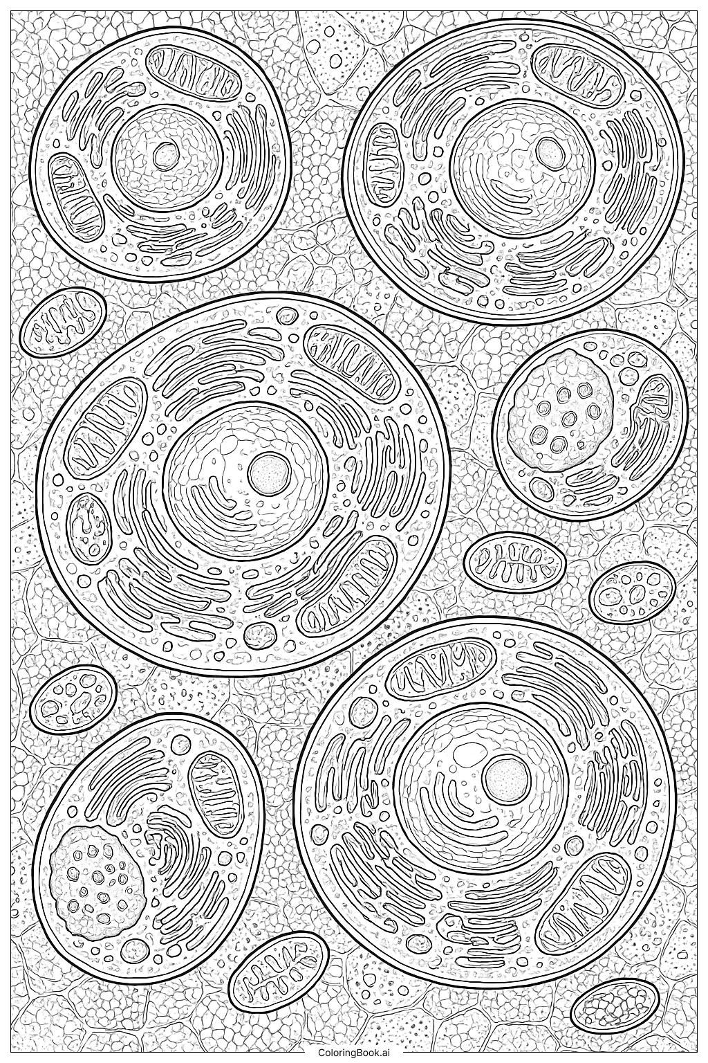
Coloring tips: How to color Cellular Level Anatomy coloring page well?
Use different bright colors for various parts of the cell to make them stand out. For example, color the nucleus in purple or blue, mitochondria in orange or red, and endoplasmic reticulum in green or yellow. The background cells can be colored in soft pastel shades to keep the main cells prominent. You can also use shading to add depth and make the organelles look three-dimensional. Feel free to use patterns or dots inside the organelles for added texture and fun.
Coloring challenges: Which parts are difficult to color and need attention for Cellular Level Anatomy coloring page?
1. The image has many small and detailed parts within each cell, which may require careful coloring and patience.
2. Differentiating between organelles can be challenging due to their similar shapes and close proximity.
3. Coloring the background cells without overpowering the main cells needs a gentle touch and soft color choices.
4. Some organelles have complex, layered structures that can be tricky to color neatly.
5. Maintaining clean edges while coloring tight spaces demands precision and steady hand control.
Benefits of coloring books: Advantages of drawing Cellular Level Anatomy coloring page
Coloring this cellular anatomy image helps children learn about cell parts in a fun way. It improves their attention to detail and fine motor skills by requiring accurate coloring of small areas. The activity also encourages curiosity about how living things work at the tiny, invisible level. Kids develop patience and focus, as the detailed design needs careful work. Overall, it blends creativity with science, making learning interactive and enjoyable.